Background
According to the Medscape National Physician Burnout and Suicide Report 2021, 36% of radiologists report burn out (some reports of burnout started before the pandemic).† To meet workload demands, radiologists need to review an image once every three to four seconds.‡ This workload will only increase as CT and MRI technologies improve in sensitivity, thereby increasing the number of images captured per patient per diagnostic test.
The adoption of AI-enabled diagnostic imaging interpretation via computer vision can help address this workload challenge by:
- Improving clinician productivity
- Augmenting imaging interpretation
- Increasing departmental throughput
- Assisting in anomaly detection, differential diagnosis, and worklist prioritization
Standard medical devices that use AI models require processing at higher frames per second (FPS) to operate effectively. GPUs are a natural choice for these models to achieve a higher FPS rate, but they are also expensive. The ability to deploy a CPU to achieve higher frames per second (FPS) and a smaller model size provides a more cost-effective alternative for deploying AI models into production.
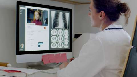
The Medical Imaging Diagnostics reference kit benefits radiology departments by using computer vision models to uncover anomalies in chest X-rays, such as pneumonia. Computer vision models are often embedded in clinical decision support systems to augment the work of a radiologist. The ultimate goal of using AI within healthcare is to achieve better patient outcomes while integrating into existing systems of the clinician’s workflow.
What Is Included
In collaboration with Accenture*, Intel developed an AI reference kit to classify chest X-rays as either normal or showing pneumonia. Each reference kit includes:
- Training data
- An open source, trained, convolutional neural network (CNN) model for image classification
- Libraries
- User guides
- oneAPI components
At a Glance
- Industry: Healthcare
- Task: Image classification to distinguish between two classes (normal and pneumonia)
- Dataset:
- 5,856 chest X-ray images with pneumonia and normal images
- 300 x 300 JPEG format
- 90:10 split (training:validation)
- Type of Learning: Supervised
- Models: CNN with 6 convolutional layers, 5 dense layers, and 6 max pooling layers
- Output: Classification of an image as either pneumonia or normal
- Intel® AI Portfolio
- Intel® Optimization for TensorFlow*
- Intel® Neural Compressor
- Intel® Distribution of OpenVINO™ toolkit
Testing Environment
Optimized with Intel oneAPI for Better Performance
Performance was tested on Microsoft Azure* Standard_D8_V5 using 3rd generation Intel® Xeon® processors to optimize the kit.
Benefits
AI medical diagnostic aids typically run multiple deep learning models in concurrency, based on the diagnostic task at hand. With this reference kit, data scientists can manage outcomes more accurately using faster FPS on a lower memory footprint due to compression of the models. This ability can save them significant costs and help to maintain or increase the value of their solutions.
Incorporating AI-enabled diagnostic imaging interpretation into a clinician’s workflow can reduce the time spent on prioritizing caseloads. The clinician can instead focus on high-risk cases requiring immediate attention. For possible high-risk patients, these AI algorithms automatically prioritize the scans in the radiologist’s workflow, which promotes faster diagnosis and quicker intervention for a higher likelihood of a better patient outcome.
References
‡ McDonald, R. J., Schwartz, K. M., Eckel, L. J., Diehn, F. E., Hunt, C. H., Bartholmai, B. J., Erickson, B. J., & Kallmes, D. F. (2015). "The Effects of Changes in Utilization and Technological Advancements of Cross-Sectional Imaging on Radiologist Workload." Academic Radiology, 22(9), 1191–1198. https://doi.org/10.1016/j.acra.2015.05.007
